
It feels like we just started this program and we’re already wrapping up. This week was the most challenging for me. Our reading for this week had less of a learning curve and I was able to understand most of it as I was reading (unlike the readings last week lol.) It pretty much covered Gaze360, a gaze-tracking dataset and method for 3D gaze estimation. It covered how they gathered the dataset using a 360 degree camera (with the top turned off) and took it to gather the data in outdoor locations. I was surprised at how small of a dataset it was with only 238 subjects, but also how effective it was. I was under the impression that machine learning datasets had to be in the thousands.
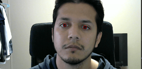
For our coding project we had to track our eye using python. According to Anil this is the first step in gaze detection and it makes perfect sense. Can’t see where the eyes are looking if you can’t accurately tack the eyes themselves. We followed a blog post by Stepan Filonov and while his explanations and procedure were easy to follow at first he kept changing the variable names so you couldn’t just copy and paste his code. I’m not sure if this was done on purpose or not? Also towards the end it seemed like it was more rushed and the new code wasn’t specified where it should go. Either way with some debugging and retracing I was able to begin tracking my eyes. Although I had to adjust the threshold first. According to Stepan:
The way binary thresholding works is that each pixel on a grayscale image has a value ranging from 0 to 255 that stands for its color. You pass a threshold value to the function and it makes every pixel below the value 0 and every pixel above the value the value that you pass next, we pass 255 so it’s white.
after changing the threshold to 51. I was able to tracking my eyes with acceptable results.
We also had a presentation by Mary! She told us about RAPID: Real-time Artificial-intelligence enabled pathology for intraoperative margin detection. basically shes going to save the world! The problem is that when doctors are evaluating tumors doctors have to make a cross-section of the tissue, stain them, and the analyze them. This is a process that apparently takes day. But with mary’s microscope they can scan in under around 10 minutes and accurately analyze the nuclei and look for anything that’s too big, dense, or erroneous.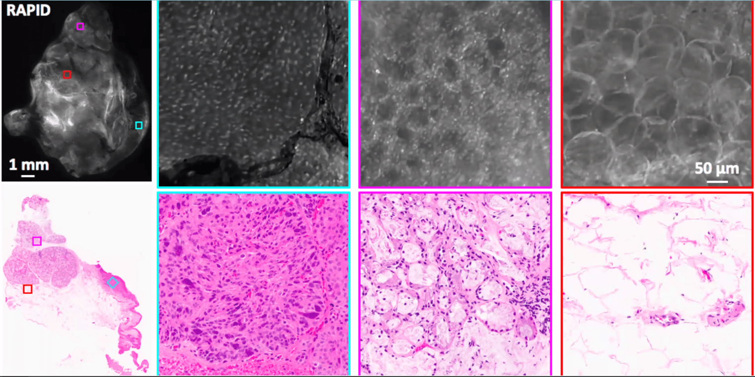
2 Responses to Week 4